Herzliya Medical Center
Tel: +972-9-959-4888
09:00-18:00
 and the fragmentation of kidney stones
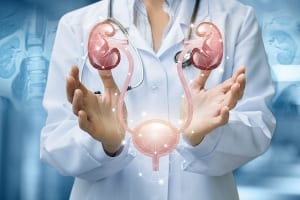
Ureteroscopy is a procedure enabling the assessment of the upper urinary tract as well as its treatment. The upper urinary tract includes two main structures that are assessed in this procedure: the kidneys and the ureters – the tubes through which urine passes from the kidneys to the urinary bladder.
Ureteroscopy is performed in instances where it is suspected that stones or malignant tumors have developed in the upper urinary tract, or when findings that have been located in it need to be treated. This procedure also enables the discovery of possible causes for bleeding in the urinary system and other problems apparently originating in this region. Ureteroscopy is performed using a catheter just a few millimeters in diameter that is introduced to the body via the urethra, usually due to the occurrence of symptoms that are suspicious for or could indicate a problem in the region: pain or burning while urinating, urgent and recurrent sensation of wanting to urinate, and others.
Ureteroscopy enables a number of procedures to be performed: Firstly, it enables a diagnostic examination, for a comprehensive, close-up assessment of the upper urinary tract, with the objective of locating various findings or causes responsible for various symptoms that have developed. Secondly, it enables therapeutic procedures to be performed, such as fragmentation of stones that have developed in this region, and their removal, removal of small tumors (including malignant tumors), and the taking of specimens from larger tumors (biopsy).
Ureteroscopy is performed under general or local anesthesia, depending on the case and the patient. The catheter used in the procedure, the ureteroscope, includes an optical system that transmits the image “seen” by the catheter directly to a screen located in from of the physician’s eyes. The physician is thus able to assess from close up the structures of the upper urinary tract, and also to specifically treat findings that require treatment.
Incidentally, ureteroscopy will not always be the recommended mode of treatment if some findings are discovered, or when the presence of interfering factors in the upper urinary tract is suspected. Sometimes, it is possible to perform treatment using shock waves, a non-invasive treatment that also enables the fragmentation of stones in the upper urinary tract.
If the procedure is performed under general anesthesia, you will be asked to fast for several hours before it. In all events, you will be asked to carry out a urine culture prior to the procedure. If you take medicines on a regular basis, please inform the staff before undergoing the procedure.
Ureteroscopy is performed, as mentioned above, by introducing a catheter with a camera at its tip, enabling the path that it traverses to be “broadcast” to the physician’s eyes in real-time. The catheter is introduced into the body via the urethra, making it unnecessary to make a surgical incision in order to access the upper urinary tract.
Ureteroscopy may begin as a diagnostic procedure and be transformed into a therapeutic procedure, depending on the findings discovered during its course. If stones are found in the upper urinary tract, they can be fragmented using a laser. The laser is introduced via the catheter to the point where the stones were found and breaks them up into small fragments. The physician will then decide whether to remove the fragments himself or to allow them to be discharged naturally.
If tumors are found in the upper urinary tract, they can also be removed, even if they are malignant tumors – but only if they are small tumors. If the tumors are relatively large, a specimen can be taken from them during the ureteroscopy, but the operation to remove them – if this is determined to be the treatment that is required – will be done in another way.
Ureteroscopy usually takes up to 90 minutes. Recovery from ureteroscopy is relatively easy: sometimes you would be released from the hospital on the day of the operation, or at the most, some two days after the procedure is completed. You will be able to rapidly resume your routine activities (in accordance with the instructions you receive from the medical staff), and you will be asked to make sure to drink plenty of fluids and urinate often during the initial days after the procedure.
The main side effects of ureteroscopy are typically burning and pain when urinating, as well as feeling an urge to urinate and the appearance of blood in the urine. These are familiar and known effects, which should pass within a few days. Infection, acute bleeding, damage to the inside of the ureter and the formation of edema in the ureter are also considered to be possible risks, but they are rare and can be effectively treated using an internal catheter introduced to the injured location and left there until the effect passes.