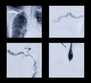
Angiography in Israel is performed in the Department of Diagnostics at Herzliya Medical Center, a leading private hospital using the most modern medical techniques and advanced technology.
What is Angiography?

Angiography is a method of X-ray examination of the blood vessels. During angiographic procedures, contrast agents are used in imaging to obtain a clear representation of blood vessels and soft tissue. The contrast agent is injected intravenously and carried through the bloodstream, to all the branches of the vascular tree. A chest X-ray is done to determine the degree of blood flow, as well as identification of any structural changes visible on walls of large veins and arteries, such as aneurysms- bulging or narrowing that causes blood vessel obstruction.
Angiography has been utilized for many years, although performed less frequently until recent medical advances have helped to eliminate side effects requiring extended recovery time. At Herzliya Medical Center in Israel, medical experts have successfully managed the surgical process to minimize side effects of angiography by the implementation of the following:
- Use of low-dose protocols
- Use of newer generation contrast agents have been engineered that reduces allergy risk and nephrotoxicity (damage to the kidneys due to a certain drug)
Why Choose Angiography at HMC?
- Utilization of modern methods for instrumental research to determine the health status of blood vessels of the heart, lungs, brain, abdominal organs, and limbs
- Highly sensitive digital devices are used to achieve high-quality images of blood vessels using low doses of ionizing radiation
- Minimized risk of side effects
- Procedure performed by leading radiologists and vascular surgeons in Israel
- Patient safety and strict compliance with international standards upheld throughout the process of treatment for each patient
The Angiogram Procedure at HMC
- Before the angiography procedure, patients undergo blood tests to diagnose renal function. From the results of renal testing, clinicians determine the necessary steps to further prepare for angiography, which may consist of circulating intravenous fluids, antihistamines, corticosteroids, or particular anti-oxidants
- On the day of the procedure, the patient must refrain from the consumption of food or any liquids (usually within 6-8 hours before the procedure)
- A catheter is introduced intravenously, through which the contrast agent is injected. For some patients, the agent is administered in a peripheral direction, though for certain procedures of angiography, the agent must remain concentrated in the immediate vicinity of the organ under investigation
- After injection of the drug, an X-ray is facilitated and processed digitally for analysis of the images generated through screening
- After the procedure is performed to produce X-ray images by way of introducing the contrast agent into a peripheral vein, patients can immediately return to normal life
Types of Angiography
Various types of angiography are used to diagnose diseases associated with vascular changes. Among the main types of angiography are the following:
- Coronary angiography – the study of blood vessels of the heart for which a catheter is inserted into the coronary artery. X-ray scanning is performed immediately after the administration of contrast medium to determine and diagnose coronary artery occlusion. Occlusion of the arteries may indicate the need for immediate, though minimally invasive endovascular procedures to resolve the blockage, such as balloon dilation and/or stenting of the coronary arteries, in order to make it possible to maintain the functionality of a blood vessel catheter. The second study helps professionals to evaluate the effectiveness of the intervention. Coronary angiography clearly show the restoration of blood flow in vascular injury after intravascular procedures
- Aortic angiography (aortography) – Process for diagnosing structural changes and allows clinicians to measure the diameter of vessel walls, including determination of the structure of the body’s largest artery
Angiography of the brain (cerebral angiography) – determination of damage usually incurred as a result of cerebrovascular accident, or as a consequence of thrombosis, embolism, spasm, vessel rupture, or congenital vascular malformation - Pulmonary angiography – an important method in the diagnosis of pulmonary circulation disorders, particularly that of pulmonary embolism. This method is also effective in the diagnosis of lung cancer, by identifying neoangiogenesis (extensive development of tumor vasculature)
- Angiography of the limbs – an indispensable method for the diagnosis of peripheral vascular occlusions. Similar to coronary angiographic procedures, this process allows immediate determinations to be made for those in need of an endovascular procedure to restore blood flow throughout the body
CT Angiography
An important advantage of using CT scanning is to perform analysis from layered images of organs and tissues. Screening generates three-dimensional computer models to help determine the precise location of the source of blood vessel functioning deficits and the effect on surrounding tissue. These essential data help plan possible surgical and minimally invasive endovascular procedures to serve the patient. Similar to other types of angiography, CT angiography is performed following intravenous injection of a contrast medium. The high accuracy of this method often allows specialists to avoid more invasive diagnostic procedures. For example, cardiac CT angiography (virtual coronary catheterization) is known as an alternative procedure to traditional coronary artery catheterization processes.
MRI Angiography
Unlike X-ray imaging techniques used for diagnostic purposes, MRI imaging is performed using a highly sensitive scanner to determine the health status of blood vessels generated by a powerful electromagnetic field. This method provides clear layered images of the highest resolution possible-essential in the process of diagnosis of small-sized obstructions and changes of blood vessels.
At Herzliya Medical Center in Israel, modern techniques for diagnosis most commonly involve MRI angiography and cerebral angiography.
 Patients Log In


